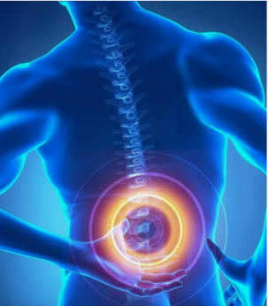
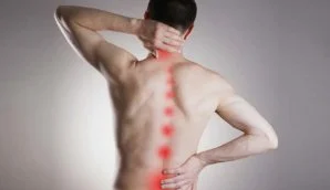
The pathology has certain features that are significantly different from those that are characteristic of the breakdown of bone tissue in individual zones and departments. The symptoms of common osteochondrosis, which characterize the overall clinical picture, are as follows:
- Significant severe deterioration in vision and hearing;
- dizziness, lack of coordination of movements;
- diseases of the oral cavity, tooth loss, skin reddening;
- General weakness, rapid fatigue of the body, even with the slightest physical exertion;
- Pain in the chest, under the shoulder blade, in the shoulder joints radiating to the upper limbs.
Widespread osteochondrosis can be localized in one area of the spine and affect multiple joints and cartilage. The signs of such pathologists are individual and clearly characterize the affected area. For example, a disease in the chest area has pronounced symptoms:
- pain and shortness of breath in the area of the heart associated with the development of intercostal neuralgia;
- Restrictions on neck and arm movements;
- Severe pain with sudden movements of the arms and trunk, sneezing and coughing;
- Diseases of the internal organs.
Symptoms of generalized lumbar osteochondrosis:
- numbness in the lower limbs and in the lumbar region;
- Pain in the pelvic area that extends to the buttocks and legs;
- seizures in the area of the osteochondrosis localization;
- Diseases of the internal organs of the pelvis associated with difficulty urinating, difficulty in defecating or, conversely, with incontinence of urine and feces.
Considering that widespread osteochondrosis of the spine manifests itself in different ways in each individual case, provoking exacerbations associated not only with the skeletal system, but also with internal organs, only a specialist can diagnose the disease. During the examination, the doctor will definitely prescribe an MRI scan, which will reveal pathological processes throughout the spine and prescribe effective treatment.
Common osteochondrosis of the spine: treatment
Treatment of the common osteochondrosis is conservative. In addition to the qualifications of the doctor, the patient himself plays an important role in the treatment. More precisely, his efforts. On the basis of the area of localization of degenerative-dystrophic changes, therapeutic measures are planned. Since the localization of the pathological process is expanded, you need to understand that the treatment will be long.
The complex of therapeutic measures includes manual therapy, physiotherapy, acupuncture, therapeutic exercises and pharmacological treatment.
Therapeutic exercises for advanced osteochondrosis or exercise therapy are an important part of successful treatment. However, this is only required under the supervision of a qualified instructor. Movement should be avoided during exacerbations as complications can arise. Consider some exercises to help relieve muscle tension:
- While standing, tilt your head to the right and left and hold it down for 5-10 seconds. Repeat this 10 times.
- Move your shoulders back and forth in a circular motion. Repeat 7-10 times.
- Make a few bends in the left and right torso.
There are also extremely severe cases, of which about 7%. They may require surgery. Surgical treatment of osteochondrosis is traumatic and difficult. Even a highly qualified surgeon cannot be a complete guarantee of success in this case. So don't take the disease to the extreme. At the first signs, you should immediately consult a doctor and follow all his recommendations, change your lifestyle and take preventive measures.
Sacral osteochondrosis and its symptoms

Sacral osteochondrosis causes complaints in the lower back and in the sacrum in the direction of the sciatic nerve. At the same time, the temperature of the lower extremities and their sensitivity decrease. Sometimes with sacral osteochondrosis what is known as "radicular pain" occurs, ie a kind of "lumbago" in the patient's leg from the hip to the tips of the toes.
A symptom of sacral osteochondrosis is also pain in the back of the surface of the lower extremity, radiating to the heel of this leg and along the outer edge of the foot, while the effect of numbness is also noted there. Well, due to disturbances in the vessels in the lower leg, the effect of cold and cold snap occurs.
In addition to the symptoms of sacral osteochondrosis already mentioned, many patients develop various disorders of motor functions, that is, paresis of some muscle groups in the lower leg, and the Achilles reflex may be absent on the side of the disease.
The examination also shows the following known symptoms of sacral osteochondrosis:
- When the patient's extended leg rises, there is increased pain in the lumbar region, in the buttocks and also on the back of the thigh from the side of the lesion - that is, the Lasegue symptom;
- with a sharp inclination of the patient's head forward, the pain in the leg and lower back intensifies - Neri's symptom;
- The moment the leg takes a sitting position lying on its back, the leg on the side of the lesion with the disease experiences a flexion reflex, that is, ankylosing spondylitis;
- When coughing, sneezing or other stress, increasing pain in the lumbar spine occurs - the so-called Dejerine symptom;
- When examined, the effect of smoothing the wrinkles on the gluteal folds from the side of the affliction is a bonnet symptom.
General guidelines for back health.
- Diet against osteochondrosis. Nutritional therapy supplies the body with the substances it needs to restore damaged cartilage tissue. There are no specific prohibitions for products. You should reduce your consumption of fatty, spicy, salty and smoked foods, and increase your fruits and vegetables. The main requirement for proper nutrition is the variety and balance of dishes.

- exercise. Regularly performing individually selected exercises according to the level of damage and the stage of the disease will help alleviate the condition and stop the development of the pathological process. At the onset of the disease, remedial gymnastics can accelerate the recovery of damaged cartilage, which can lead to complete recovery.
To keep the body in good physical condition, you can choose any direction of sports: running, swimming in a pool, Biking, running, skiing and inline skating, dancing, yoga, qigong.
When choosing your sport, you should consider the effects of the activity on the body. - Hypothermia and stressful situations should be avoided.
- Appropriate load on the spine. Sharp turns, slopes, lifting and carrying weights should be avoided. When heavy work is required, safety precautions must be taken. Monitor your back. If there is an overload in a certain muscle group, it is necessary to take measures to relax. Sedentary work is the same. Regardless of the type of activity, you should alternate between rest and work. Throughout the day, regardless of the type of activity, monitor the correct position of the body in space, its straight posture and compliance with biomechanics when lifting weights. These simple rules of conduct require human attention, self-control is required at all times.
- Corsets should be worn with spinal diseases, especially during strenuous physical activity.
- Quit alcohol and smoke.
- Correctly coordinated sleeping equipment: firm pillow, medium-hard mattress, bed with a firm sleeping base.
- Wear comfortable shoes. The love of fair sex with high heels has a detrimental effect on the condition of the spine, especially the lumbar spine.
- body hardening.
- Massage and self-massage.
Following simple rules avoids many diseases, which significantly improves the quality of life. A healthy back is based on exercise and proper nutrition. Overloading the spine is more common in a sedentary lifestyle than in physical work. If it is not possible to get rid of harmful factors that lead to the destruction of the cartilage tissue of the intervertebral discs, then the body should be trained to increase resistance to their influence.
Use physiotherapy
Electrophoresis and medication:
Electrical impulses help the medication reach the target area faster. This type of physiotherapy is done with aminophylline or novocaine, with the first improving blood circulation and the second reducing pain.
Ultrasonic effect:
Stimulates the metabolism and related processes. Relieves pain, relieves inflammation.
Magnetotherapeutic effect:
Helps with edema, relieves pain.
Laser therapy:
Helps with inflammation and improves the functions of the circulatory system.
Why is the problem developing?
Several different reasons are characteristic at the same time for widespread osteochondrosis of the thoracic spine, as well as for a similar pathology in other areas. For example, rheumatoid factor is said to be the most common catalyst. According to statistics, around one third of patients suffer from arthritis of large joints at the same time.
Also often causes symptoms of such a serious problem and osteochondrosis of part of the spine, because, due to this, various structural changes begin in adjacent segments. In addition, it should be understood that the cause of the development of the pathology can easily become an unhealthy diet and the associated obesity. It is difficult for the spine to carry excess weight, which starts its destruction.
Often the development of such a problem is attributed to problems with metabolic processes in the body. In this case, the blood circulation slows down, regeneration deteriorates, etc.
The list of "provocateurs" can also contain:
- Inadequate and improper development of the ligaments and muscles that surround the vertebrae: this problem often manifests itself in a sedentary lifestyle
- suffered various types of injuries and operations
- Excessive physical activity: Those that last longer are particularly dangerous; and we talk about both athletic and professional loads

Prevention methods
The most effective way to deal with osteochondrosis is with proper diet and exercise. With osteochondrosis of the spine and treatment with gymnastics, the following have shown the greatest efficiency:
- Massage of the lumbar spine, back and limbs in the morning;
- jumping in place, exercises on the horizontal bar;
- Regular breaks for physical education classes during work, 7-9 exercises are enough to prevent the disease;
- Swimming, especially recommended by back stroke specialists.
Most patients consider how to begin treatment for back osteochondrosis when signs of osteochondrosis appear and intervertebral osteochondrosis is already progressing. In this case, the general recommendations from the complex of physiotherapeutic exercises for stretching the spine should be given.
If osteochondrosis occurs, it is necessary to adhere to a strictly balanced diet, which is mainly based on protein feed, but excluding fungi from the daily range as much as possible. You should limit yourself to eating salty, fatty foods, including homemade pickles. Sugar, flour, and confectionery are also contraindicated.
Bad habits have to be given up in order not to consume too much coffee and products based on it. The daily volume of fluids you drink should not exceed 1 liter, and the number of meals should be between 5 and 7 per day.
The lack of vitamins and nutrients, which has been proven by analysis, requires their immediate replenishment in the body. This requires the intake of multivitamin complexes.
gymnastics & massage
Exercise therapy in the treatment of osteochondrosis plays a leading role. Without exercise therapy, it is impossible to form a strong muscular corset, and the latter is urgently needed to maintain a diseased spine. Gymnastics also increases blood circulation in the vertebral zone, improves metabolic processes and helps to quickly remove caries products.
How do you cure osteochondrosis with gymnastics? The complex is selected only individually and can only be carried out in 1-2 steps without medical supervision. In later stages, unnecessary, vigorous movements can shift the disc and increase the problem. In stage 3, all exercises are only performed in the prone position.
For the treatment of osteochondrosis, massage is a must. In the acute stage, it is not done - it will cause a thrill. However, properly performed massage at the chronic stage with osteochondrosis is irreplaceable. After a session, the muscles relax, the braces are removed, and nerves and blood vessels begin to function normally. The massage is performed only gently and without sudden movements. You cannot entrust your spine to a layperson!
Prevention of sacral osteochondrosis
It should be noted that it is almost impossible to completely get rid of the disease with sacral osteochondrosis. The level of development of modern medicine can only help to prevent the further development of this disease.
In order to prevent sacral osteochondrosis of the spine, it is necessary to start maintaining the normal condition at a young age. To do this, try more often to at least hang on the crossbar. This way, you remove the load from the spine and help it relax.
Since sleep makes up a third of our lives, it is important that the spine is in the correct position while we sleep. Therefore, the best sleeping position is on your back.
Well, of course, a balanced diet high in dairy and seafood, as well as legumes, helps control the body's metabolism.
Symptoms of cervical osteochondrosis
The neck region contains many blood vessels that intensively nourish the brain. Therefore, the risk of osteochondrosis is associated with a poor supply of blood to the organs of the head, especially the brain. The signs of osteochondrosis of the cervical spine depend on the segment affected. Therefore, the following signs of cervical osteochondrosis are distinguished:
- radicular syndromes;
- cardinal syndrome;
- stimulus reflex syndrome;
- vertebral artery syndrome;
- Compression of the spinal cord.
Pain sensations occur in the neck, move into the zone of the shoulder blades and can reach over the shoulder and forearm to the fingers. The characteristic signs of cervical osteochondrosis in this case are tingling, burning, pâté, numbness of the hands, forearm or fingers.
Osteochondrosis of the cervical spine has the following symptoms in irriative reflex syndrome. The main symptom is the presence of acute, intense, burning pain in the cervico-occipital area or in the neck itself. The pain tends to increase with movement or exertion, it is especially acute after a static state. For example, after sleeping with inaccurate head turns after sleeping. Symptoms of cervical-thoracic osteochondrosis show a wider spectrum, the most basic of which is disruption of internal organs.
Vertebral Artery Syndrome - exacerbation of cervical osteochondrosis while symptoms are extremely intense and consist of severe, burning pain. Characterized by throbbing pain in the temples, the presence of severe migraines, particularly affecting the parietal part, the back of the head and the forehead region. The pain is chronic, constant, but sometimes cases of paroxysmal pain are recorded. Worsening osteochondrosis of the cervical spine expresses symptoms in the form of increasing pain. This happens after uncomfortable positions, sudden movements and heavy physical exertion. An exacerbation threatens hearing impairment - a disorder of the vestibular apparatus, the experience of tinnitus, as well as a decrease in hearing acuity. Vision problems are common.
Osteochondrosis of the symptoms of the cervical spine in cardinal syndrome. Severe angina is characteristic of cardinal syndrome. It makes it difficult to identify the underlying disease - osteochondrosis.
The phenomenon of angina pectoris is caused by pressing the root in the lower segments of the neck region, which finds a reflex response. Cardinal syndrome can manifest itself in cases when the roots of the pectoral muscle or the phrenic nerve are irritated. Osteochondrosis of the cervical spine, symptoms of cardinal syndrome can manifest themselves in the form of tachycardia, extrasystole. There are common cases when osteochondrosis of the cervical spine and high blood pressure occur at the same time.
reasons for
After numerous in-depth studies, experts have come to the conclusion that the main reason for the occurrence of osteochondrosis is the uneven distribution of load on different parts of the spine. The main cause can be intensive work in unusual conditions, long sitting in one position, heavy loads during sports training, strikes. The result of unexpected or prolonged stress on a certain part of the spine is a gradual change in the structure of the intervertebral discs.
Among the main reasons for the onset and gradual development of the disease, several main reasons can be distinguished:
- Hereditary changes in the development of the body that lead to osteochondrosis.
- diseases of the endocrine system, malfunction of metabolic processes.
- Abnormal development of the musculoskeletal system leading to pathological changes in the body.
- Injuries to the back, lower back, neck during falls, training, sudden loads, bumps.
- An inactive lifestyle burdened by poor nutrition.
- Osteochondrosis is a constant companion of overweight people who are overweight.
- Alcohol and nicotine abuse inevitably leads to the destruction of the intervertebral discs.
- Constant psychological and physical stress, stress and overload become the main cause of the dystrophy of the intervertebral cartilage.
- Women at various stages of pregnancy are often faced with manifestations of osteochondrosis.
People of some professions are most susceptible to osteochondrosis, since the monotonous performance of their tasks gradually leads to dystrophic changes in the structure of the intervertebral cartilage. The main risk groups are:
- Accountant.
- Cashier and Manager.
- Driver of a vehicle.
- People who play sports.
It should be borne in mind that women are most often affected by the disease due to a poorly developed muscle system.
Development of osteochondrosis
No illness occurs without a cause or begins immediately. There are four main stages of the disease that you need to know in order to see a doctor on time.
- Gradually beginning dystrophic changes in the nucleus pulposus of the intervertebral cartilage usually go unnoticed. Disc dehydration becomes the main cause of micro-tears, loss of elasticity, and thinning of cartilage. Often times, people feel a little uncomfortable at this stage, sitting in one position for long periods of time, or experiencing unexpected lumbago during intense exercise.
- The second stage is hard to miss. Degenerative tissue changes lead to a head start. The fiber capsule collapses and the intervertebral space contracts. The result is pinched nerve endings, the appearance of sharp pain in certain areas of the back. The pain syndrome is actively expressed on inclines, sharp turns and running. Osteochondrosis is accompanied by a sharp loss of the ability to work and the appearance of weakness in the body.
- Complete or partial abrasion of the cartilage mucosa due to osteochondrosis. The thinning of the tissue is clearly visible during radiography. The symptoms of the disease are pronounced and can lead to partial paralysis. There is no pain relief, and you will have to resort to injections and other medical effects to determine the focus of the disease. Only strong medication and complete rest will help.
- This is the final stage, characterized by complete destruction of the intervertebral cartilage. A complex disorder of the neurological system that leads to the appearance of bone growth instead of cartilage tissue. The mobility of the joints is completely impaired. Osteophytes can injure the nerve endings of the vertebral and bone segments. At this stage, the help of a surgeon may be needed for treatment.

Diagnosis
Reference is made to family history as osteochondrosis has a genetic component. The specialist asks about the workplace, living conditions and the course of the disease itself, and the patient has to describe exactly what worries him
The best result can be achieved with good feedback between the patient and the doctor.
The next method is an objective study, carried out by the specialist himself or using instrumental methods. The doctor will check the range of motion of the neck and limbs, which may become noticeably narrower due to pain and stiffness. Palpation records how much the muscles are subjected to cramps and how curved the spine
isAttention is drawn to a neurological examination that can be used to track disturbed reflexes. This symptom can be the result of compression or damage to a nerve
hisdiagnose the disease using MRI, X-rays, myelograms, and other methods
Instrumental methods of diagnosing common osteochondrosis include:
- X-ray of the entire spine in two projections.
- MRI to assess ligaments and nerve tissue.
- Electrophysiological study to check the transmission of nerve impulses.
Radiography is effective in determining the presence of bone growth - osteophytes, narrowing of the spinal canal and the presence of other diseases that are a consequence of osteochondrosis, such as: B. Scoliosis.
Computed tomography can also be used with MRI. CT can determine the degree of compression of the nerves with spurs.
The diagnosis of common polysegmental osteochondrosis is made when other pathologies that cause vertebral destruction (e. g. tuberculosis) have been ruled out and when multiple segments of one or more departments are affected.
There are additional diagnostic methods. These include:
- bone scan.
- discography.
- myelogram.
Bone scans can detect diseases such as osteoarthritis, fractures or infections. This method is a radionuclide method and is suitable for differential diagnosis and for the identification of possible complications.
When performing the discography, a contrast agent is injected into the gelatinous (pulpous) core of the intervertebral disc. This method is effective in determining the presence of a herniated disc.
The myelogram is also a contrast study method. The contrast is injected into the spinal canal and the image is recorded using X-ray or CT. This method allows you to determine the condition of the vertebral cocoa, the presence of constrictions and bruises.


